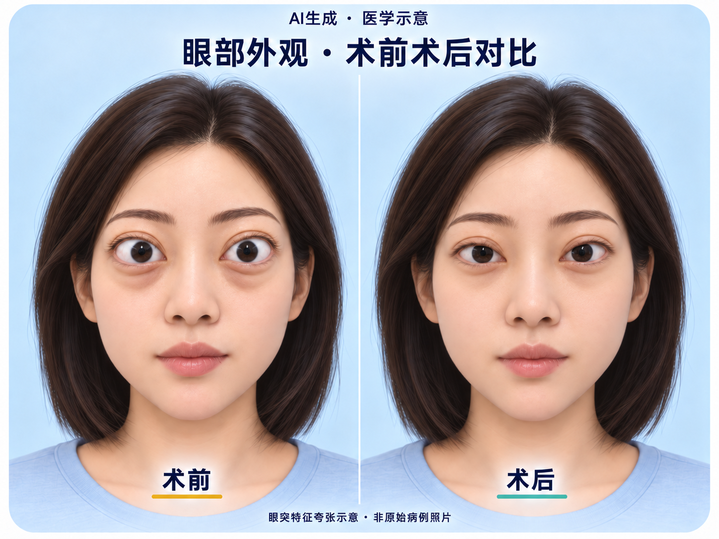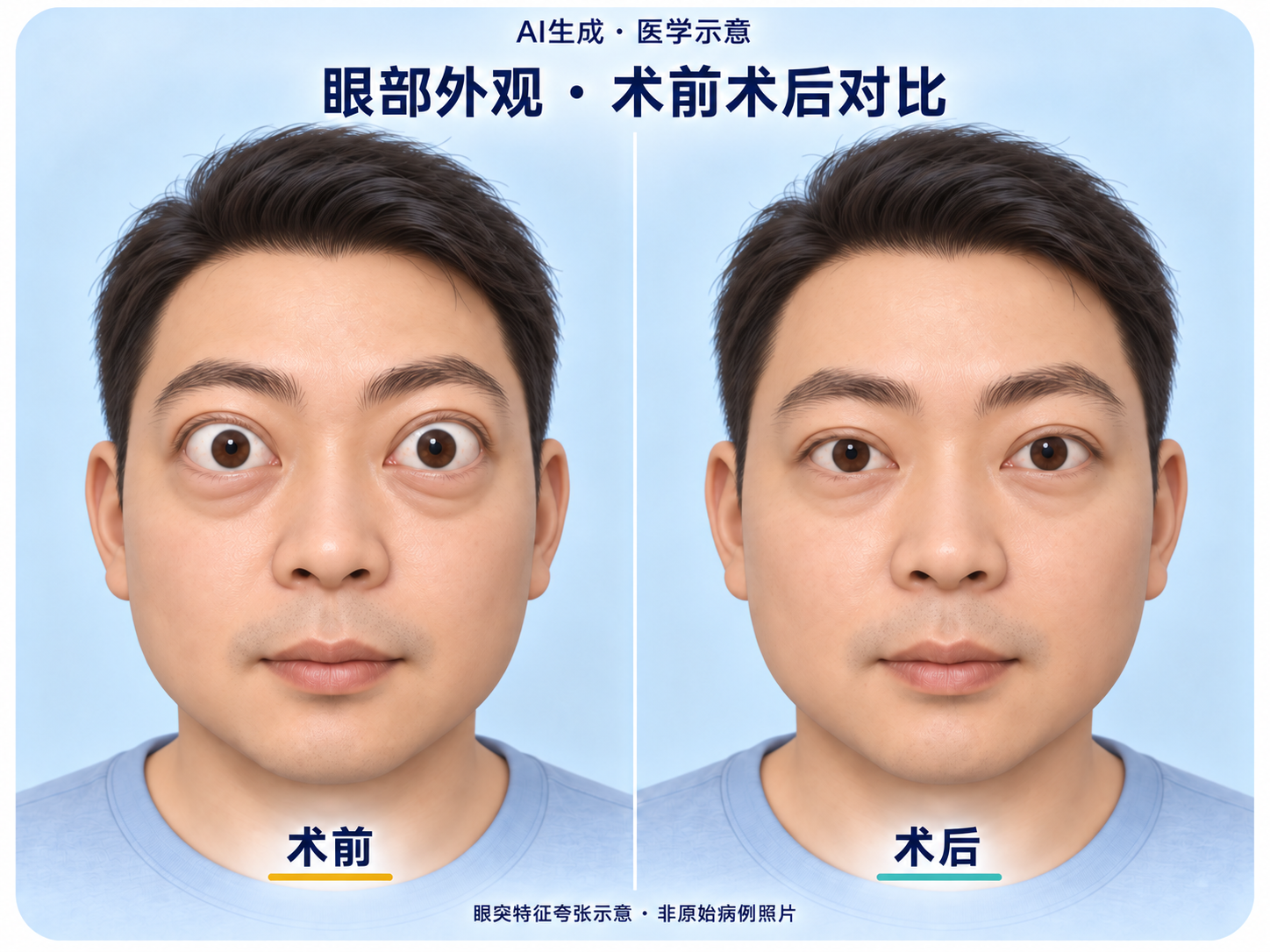
Eye appearance illustration · Set 1
Fictional people illustrate differences in eye appearance to aid understanding of medical concepts.
AI-generated · Not original patient photographs
View the full illustrationCASES & INSIGHTS
Explore changes in eye appearance and surgical principles through medical illustrations and actual clinical images.
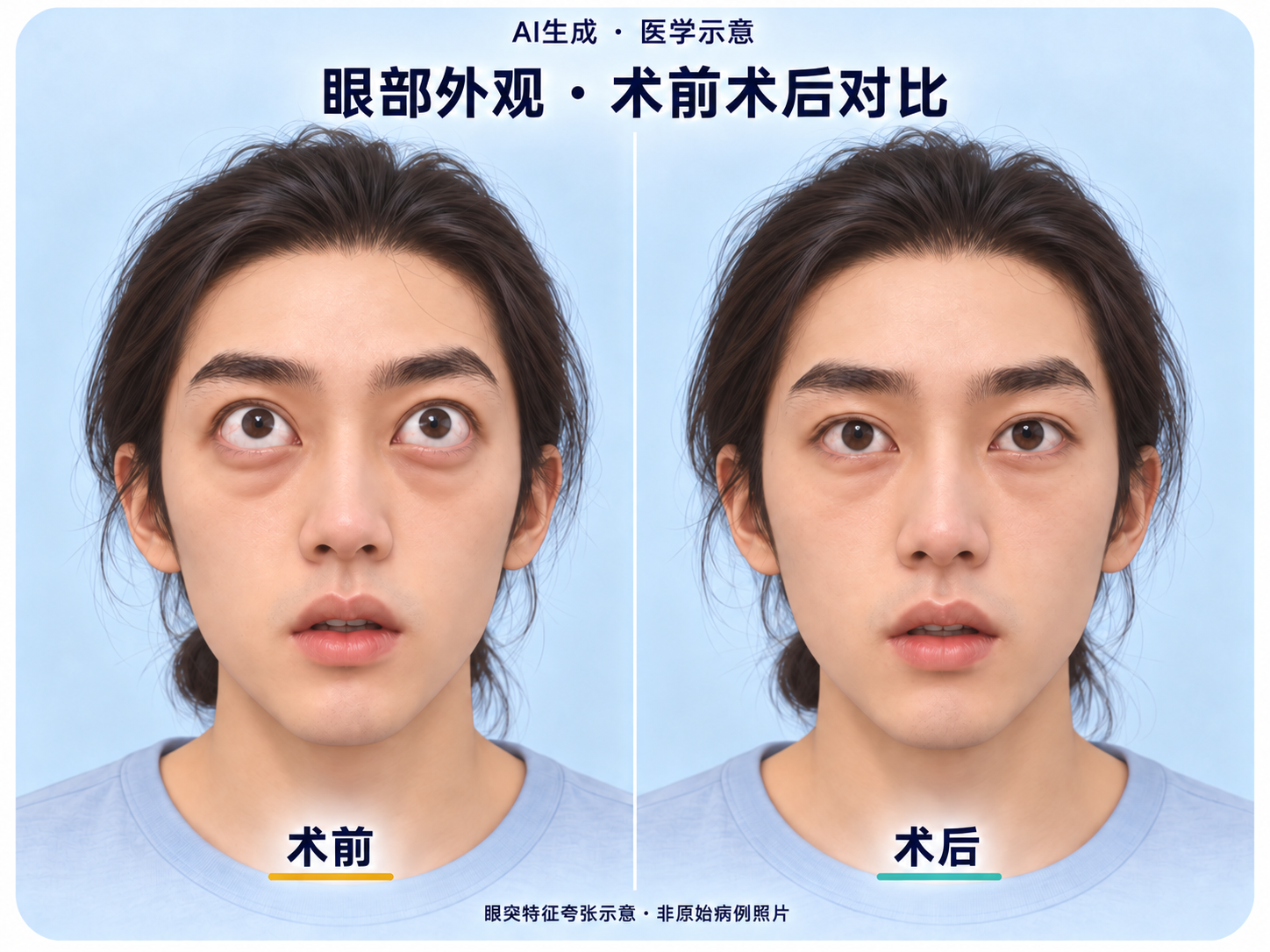
Fictional people illustrate differences in eye appearance to aid understanding of medical concepts.
AI-generated · Not original patient photographs
View the full illustrationFictional people illustrate differences in eye appearance to aid understanding of medical concepts.
AI-generated · Not original patient photographs
View the full illustrationFictional people illustrate differences in eye appearance to aid understanding of medical concepts.
AI-generated · Not original patient photographs
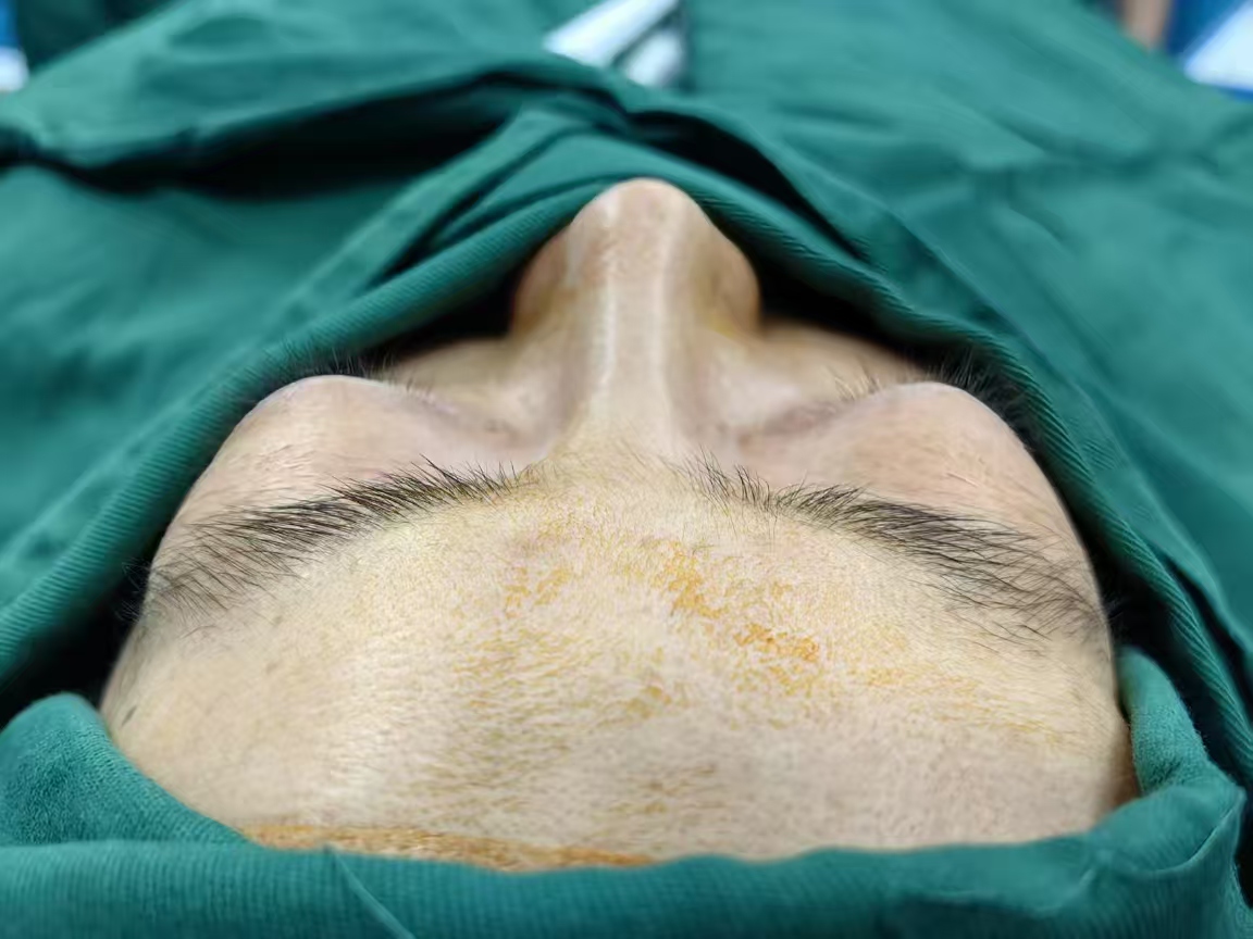
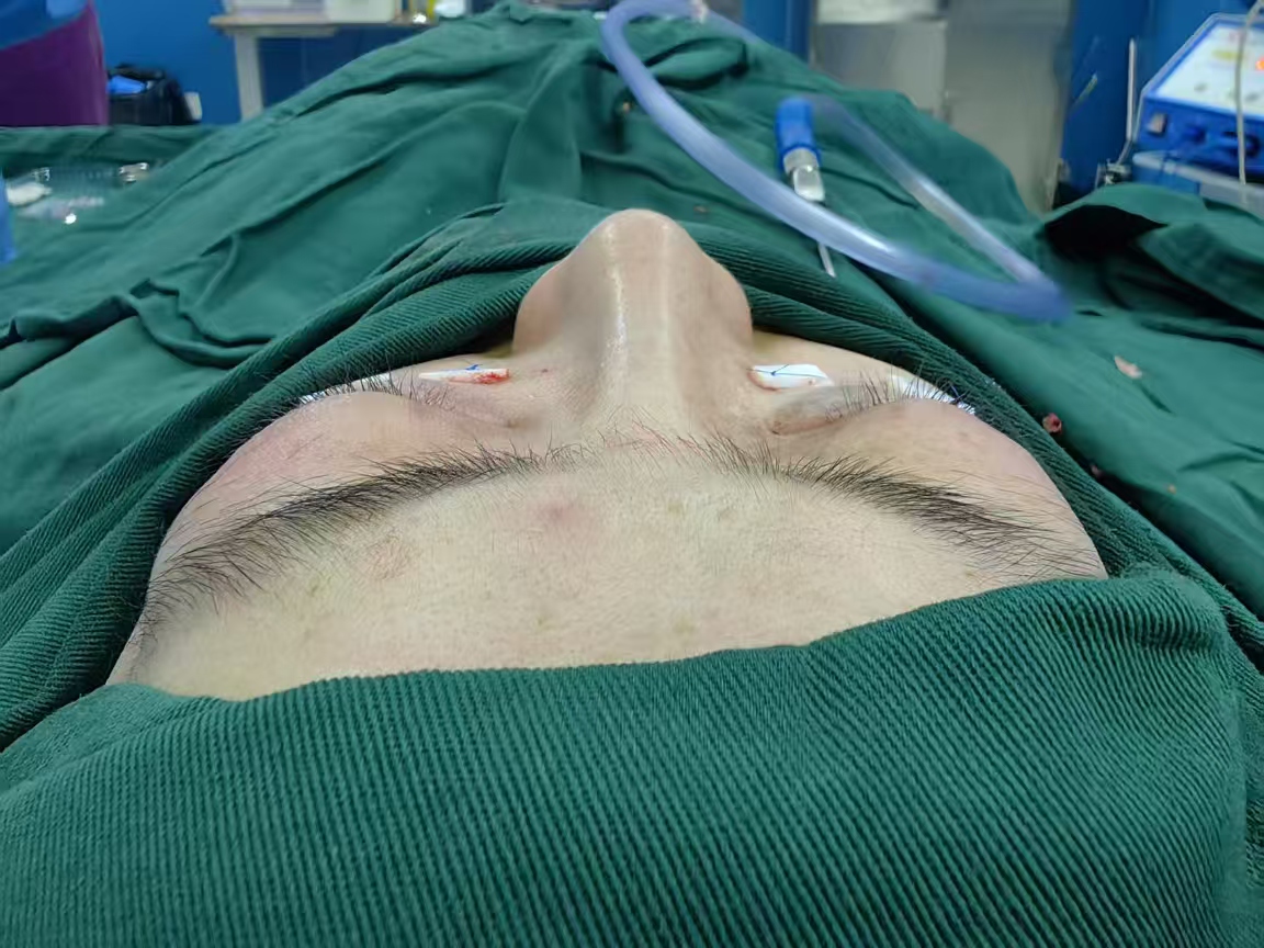
View the full illustrationPreoperative and postoperative photographs from the same case are shown side by side, preserving their original angles, proportions, and framing.
One case · 2 original photographs
View the case imagesAI-generated educational demonstration · Not an actual case · Not a prediction of outcomes
Original Chinese audio with subtitles in your selected language
How can a protruding eye gradually move back? First, look at the change in its outline, then explore the principles behind it.
Why does the eye protrude? The eyeball sits in a space enclosed by bone, called the orbit.
When fat takes up more room, or the bony walls extend farther inward, space within the orbit becomes more limited.
When this space becomes crowded, the eyeball may be pushed forward.
Where does the surgeon enter? First, the eyelid near the outer corner of the eye is retracted.
This exposes the conjunctival sac inside, where a small, concealed incision is made.
To create more space, small amounts of fat are removed from this area in stages.
This gradually reduces the volume occupied locally while preserving and protecting the surrounding tissue.
Next, look at the deep bony wall on the outer side of the eye. This is mainly the greater wing of the sphenoid bone.
Within the planned area, a portion of the bone is gradually removed with a surgical burr.
Only a localized area of deep bone is removed. The orbital rim at the front remains intact.
The thin membrane surrounding the orbital tissues is then opened as needed.
Some of the soft tissue can then expand into the additional space.
The bony framework stays in place as the space changes. With more room available, the eyeball may be able to move back to a more appropriate position.
The approach shown in this video requires no separate skin incision. The entry point is concealed on the inner surface of the eyelid.
In the approach shown here, the small incision is left unsutured. The conjunctiva heals naturally after surgery, so no stitches need to be removed.
Creating more space is the principle behind orbital decompression. Follow your doctor's advice on the specific procedure and recovery plan.
The following is a translation of the aftercare text in the original video, not a standard care plan suitable for everyone. Follow the medical instructions given for your individual case.
Apply cold compresses intermittently as instructed by your doctor, using a clean gauze barrier and avoiding pressure. Keep water away from the wound for the first 3 days.
Avoid bending over, lifting heavy objects, holding your breath, blowing your nose, and similar actions.
Gently pull down the lower eyelid to apply eye drops. Use eye drops, eye ointment, and oral medication at the doses and for the duration specified in your individual prescription.
Follow the hospital's care sheet: temporarily avoid spicy food, very hot food, and seafood. Avoid smoking and secondhand smoke.
Attend follow-up visits 1 week, 2 weeks, or about 1 month after surgery, as scheduled. Book Dr. Hui Chen's outpatient clinic in advance.
If your vision suddenly worsens, pain increases, or swelling develops rapidly, contact the surgical team or an ophthalmic emergency service immediately.
In the procedure shown in this video, the small conjunctival incision heals naturally and does not require suture removal. Follow your individual medical instructions for specific care, medication doses, and treatment duration.
Animated illustrations introduce the basic principles of orbital decompression, with visuals and narration explaining the surgical approach.
02:07 · Hui Chen team moving-watermark edition
Watch the full explanation